EMBRYOS?
THE DOUSHANTUO MICROFOSSIL ASSEMBLAGE
|
The Doushantuo 'Embryos'
Microfossils from the Doushantuo Formation were first described in ??? (REF), and described as algal fossils and acritarchs. However, a sub-set of the specimens were interpreted as the embryos of metazoans (Xiao et al., 1998); a suggestion that sparked considerable debate, since they may extend back as far as 600 million years in age. Arrangements of 1, 2, 4, 8, 16, 32 cells have now been supplemented by specimens showing all numbers of 'cells', up to 100s, some of which now occur in peanut and spiral morphologies. Alternative suggestions that the specimens represent giant sulfur bacteria (Bailey et al., 2007), and the embryos of non-metazoan holozoans (Huldtgren et al., 2011) have been debated. A host of high-resolution imaging, geochemical and petrological techniques have been used to address the question of what these specimens may be, but the most recent publications on this material are yet to reveal a clear interpretation (Chen et al., 2014; OTHER REF). |

A digital model of an embryo-like fossil from the Doushantuo biota. The structures in each cell shown in yellow have been interpreted by some palaeontologists as nuclei, though this has been controversial. Image courtesy of J. Cunningham.
|
A spiny acritarch from the Doushantuo biota imaged using synchrotron tomography. The affinities of these fossils are unknown. Image courtesy of J. Cunningham.
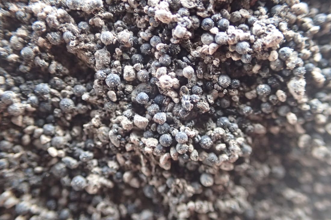
Microfossils within a phosphorite from Weng'an, South China. On closer examination under a microscope, some of these spheres may be identified as embryos. Image: A. Liu
